
When It Hurts to Say I Told You So: Advanced Prostate Cancer Cases on the Rise

emperorDiagnose a patient’s prostate cancer in its earliest stages and you have a great chance of curing it. Starting in the early 1990’s, that was the philosophy behind Prostate Specific Antigen (PSA) blood testing in men. It wasn’t a perfect test, and many men with increased PSA proved not to have cancer when their prostates were biopsies. Some other men underwent heavy duty treatment such as prostatectomy or radiation for prostate cancers that were SO small and bland they probably would never have spread. Despite the drawbacks, the death rates from prostate cancer dropped in men who were being screened.

As I have written about before. some scientific panels, as well as CMS, the federal agency that oversees Medicare, took a dim view of PSA screening, and the number of men being screened decreased. Some men were never tested, others were still getting tested, but maybe a few years later or a little less frequently than they had been in the past. As a busy prostate pathologist this delay in testing worried me, and I never missed an opportunity to tell the men I knew to have PSA testing. I told women to nag the men they love. And I never failed to have my PSA tested as part of my annual physical

My worries soon began to become reality. My colleagues and I, examining biopsies under the microscope, began to see more cases of aggressive looking (higher Gleason Grade) prostate cancer than we had in years past.  This was what is called anecdotal findings; it was our impression, but we did not do a statistical study or a publish a peer reviewed scientific paper to state our claim and have it verified (or debunked) by other pathologists around the country.  But we wondered, was there a correlation between what we were seeing and the amount of spread of cancer in the men being diagnosed?

We now have the beginning of an answer. A study published this week from my alma mater, Northwestern University, confirms that a higher number of men are once again being diagnosed with advanced prostate cancer. By the time of diagnosis, their cancer has spread to bones or lung, making cure much more unlikely. The window of catching a small, treatable, tumor has passed. The authors do not claim this is entirely due to lack of PSA screening, but to quote the lead author, Dr. Edward Shaeffer, “If I were a patient, I would want to be vigilant. I firmly believe that PSA screening and rectal exams save lives.”

PSA isn’t perfect. We hope someday to have a better test. And, very importantly, any man diagnosed with prostate cancer needs to have a long discussion with his urologist about the best way to manage his disease. But mammas, don’t let your babies grow up to be victims. Tell them PSA’s save lives.

————————————–
You never know what you will find here. To subscribe, type your email address in the box and click the “create subscription” button. It is completely spam free, and you can opt out at any time.

Reading a Prostate Biopsy-Inside the Mind of a Pathologist

tray
One Patient’s Prostate Biopsy Slides

West Suburban Chicago

6 a.m.

“Let’s see, how many prostate cases do we have today. Looks like I get nine. Better get started now before the phones start ringing and before the lab techs start popping their heads in. And oh yeah, I will have urine cytology slides and FISH heading my way.

“Too deathly silent in here though, better turn on the radio. The Sox lost last night, so no point in listening to the Mulley and Hanley on The Score. I’ll flip to FM  and go with Lin Bremer on “XRT.  A little music and a chance at ‘Three for Free.”

“Got my tea, let’s find the first case. My case list start’s with Bobby Bright, routine 12 part biopsy. That will be one of the big trays that can hold all 24 slides. Oh, here it is, top of the stack. All the slides have their labels, and the labels match the requisition and billing sheet. Good, no issues. Flip on the microscope light, grab the first slide and we are rolling.

“Part A is from the right apex.  A few inflammatory cells, but nothing that looks suspicious for cancer. Check off the benign code on my worksheet. Part B, right middle, oh-oh. I don’t like that group of glands. Their growth pattern is too irregular. Let’s look at a higher microscopic power. Yup, there are big nucleoli, and I don’t see any basal cells. Pretty sure this is malignant, but am I sure? I think I will order a PIN4 stain. The insurance company won’t be happy paying for it, but they would be more unhappy paying for a prostatectomy the patient doesn’t need. Mr. Bright wouldn’t be too happy about any of that either. So let’s do the stain. I should have it by this afternoon in time for case review, so there won’t be any delay. Rest of the case is benign, good for this guy, he might only need active surveillance, no surgery or radiation for now. Oh, I better remember to flag this case for possible molecular testing. That will help decide the treatment question.

“There’s my microphone. I guess I forgot to turn it off last night, but it’s still charged. Good thing, I can dictate the diagnoses without having to recharge. The staff can start typing early.

“OK, next case, Grant Aspen. Wow, these glands look really stunted. And the cells are so bizarre. But that blood vessel doesn’t look right either. Hmm, I think I know what’s going on, but let me check the medical chart. So glad we have the electronic health record, it really makes my life easier. Yeah, just what I thought. This guy had prostate cancer five years ago and was treated with radiation therapy. All those freakin’ changes are from the rays. No cancer here. Let’s get this dictated and move on.

“Next case is one of those MRI-Fusion biopsies. Sometimes that MRI is really good at picking out the area in the prostate where the risk of cancer is high. That will be Part M on this case. Here it is, yeah, that’s tumor for sure. But how do I want to grade this. Is this a Gleason 3+3, or a Gleason 3+4. It’s hard to tell on this level. Good thing we have 6 levels on each biopsy. This next level definitely has poorly formed glands, so we’ve got some Gleason Grade 4 here for sure. That will make the Gleason Score a 3+4=7. And I see cancer in some of his other biopsies as well. I am afraid he is going to wind up in the OR for a robotic prostatectomy. Better dictate to flag this patient for our Quality Assurance audit. After he has his prostate out  I can check that pathology report against our biopsy report.

“Damn, there goes the fire alarm. Forgot that we are having a fire drill today. Oh well, the sun is shining and a few minutes standing around in the parking lot will help clear my mind.  Gotta keep sharp, never know what the next slide will show.”

——

You never know what you will find here. To subscribe, type your email address in the box and click the “create subscription” button. It is completely spam free, and you can opt out at any time.

Standard Operating Procedures.

no countryIf the rule your followed brought you to this…

…what use was the rule?

No Country for Old Men — 2007

Standard operating procedures (SOPs) are the life blood of the laboratory. They tell us what we are doing and how to do it. The best ones even tell us why we are doing it. I would never suggest changing an SOP without validating any new process, verifying that any changes produce results at least as accurate, timely and cost effective of what came before. But rigid procedures can lead to monotony and lack of attention. And lack of attention can lead to errors. Sometimes just a tiny tweak, not in the procedure itself, but how you go about following it, can freshen up an otherwise mundane task.

In my work as a urologic anatomic pathologist, I spend several hours each day examining prostate biopsies. Now, I cannot vary that I have to look at each slide carefully, need to make decisions about presence or absence of malignancy, grade the tumors I observe, and record my findings. Those steps are immutable. And after 11 years, they could easily be cemented in stone. But I have discovered that making small changes in the way I go about each step gives me elasticity and prevents mind freeze.

What kind of changes am I talking about? First, I have to admit that there was one big one. For years I had been using the same microscope, actually one I brought with me from my old hospital position, an older ‘scope the hospital sold to me for a song. Last year my colleagues convinced me that an ergonomic microscope with a tilting head and eyepieces would go a long way toward preventing back and eye strain. And that’s important as I enter my fourth decade of peering down the ‘scope — something both my Lab Safety Officer and more importantly my Occupational Therapist wife, agree with.

So that was a pretty big variation, but most of the others have been small, simple and inexpensive. I changed the location of my microscope, putting it just a quarter rotation of my chair away from my desktop computer. As we have become integrated with an electronic medical record, I find myself more and more frequently logging into the computer to get some patient information from the EMR, so the proximity is a real time saver. And the change in “locale” made the work seem fresh again.

My latest initiative changes the way I dictate my cases. I have traditionally used dictation equipment using a foot pedal so that I can dictate in real time as I look through the ‘scope. We progressed from tape to digital a few years ago, but that didn’t change my style. However, I  recently decided to record my findings in writing and then dictate at the end of each case. I developed a worksheet that makes it simple to record Gleason Score and extent of tumor in my prostate cancer cases. It’s a snap to read my findings off the worksheet after each case. Now I have a written record in case the dictation gets disrupted. And no complaints from the transcribers so far.

Even outside the lab, “SOPs”, can be useful, but find a way to keep things fresh! It may be adding nutmeg to a recipe (easy), finding a new route to the job (moderately challenging), or building a new house (insane). It will keep you rocking.

(Note: To my colleagues on the various lab listserves, feel free to distribute)

———-

You never know what you will find here. To subscribe, type your email address in the box and click the “create subscription” button. It is completely spam free, and you can opt out at any time.

5 Things Your Pathologist Never Told You


Robert PalmerDoctor, doctor…

…give me the news.

Robert Palmer-1979

We have been called “The Doctor’s Doctor.” Pathologists stood in the background for decades, running laboratories, interpreting biopsies, staging tumors and making diagnoses. We rarely exposed ourselves directly to patients. There is even a law in New York State that explicitly prevents such contact. But now we live in a much more connected world. At the dawn of the digital age I created Pathwise, a company that “translated” biopsy reports from medical jargon to easy to understand English for patients with a desire to know. Now pathologists communicate via the Web, via Facebook, via Twitter. But even with all this improved interaction, there are still things you may not know about what really happens to your blood or your kidney or breast once it exits your doctor’s office, your hospital bed,  the O.R. And even less that you might know about the pathologist who coordinates all the activity.

  1. Most likely, the laboratory that analyzes your specimens has a pathologist as a Medical Director. To have an accredited laboratory, there is a long list of obligations and requirements that the Medical Director must ensure are occurring. This does NOT include running every blood test or making every slide personally. It DOES include verifying that qualified, well trained personnel are on hand to perform those tasks. And the scrutiny we are under to guarantee that keeps on growing.
  2. Pathologists deal with our own version of Moore’s Law. In computing, Moore’s Law predicts that over time, denser and more powerful integrated circuits will be developed. In pathology, we are faced with the proposition that tissue samples are getting smaller and smaller, but the amount of information we need to derive from them is getting greater and greater. In the last decade, tests looking at cancer cells for the various changes in their genes–how these cells differ from normal cells–are becoming essential in determining the precise type of tumor in a biopsy, as well as in deciding what chemo- or immunotherapy treatments will work best for a particular patient. And by the way, those tests are not cheap.
  3. The concept that there is a sharp line that separates “benign” from “malignant” can be a false one. Yes, in the vast majority of cases I can unequivocally call a biopsy benign or malignant. But that border line can be fuzzy; there can be a gray zone. Sometimes we just cannot accurately predict how a tumor will behave. And it is not just us local diagnosticians who are in this quandry. National experts don’t always agree on the nature of what they see under their microscope. And it can be a moving target. As new techniques are developed, and as our knowledge of the molecular underpinnings of illness grow, our interpretations and naming of disease processes change as well.
  4. You may be surprised to see a bill from a lab halfway across the country. Not all of your blood tests or tissue tests are performed locally. Some large national laboratories operate central hubs which may handle all specimens from a wide geographic area. Some more esoteric tests, particularly molecular and genetic studies on tumors, may only be performed at one laboratory in the country. Laws relating to patenting of genetic testing have changed, but laboratories can still create test combinations that are difficult to duplicate elsewhere.
  5. Pathologists are frequently leaders in a Medical Center. With offices and laboratories in the hospital, the pathologist often spends more time in the building than their clinical colleagues. This, combined with a general sense of trustworthiness, lead to many roles both within the administration of the hospital and the concerns of the physicians that make up the medical staff.

One more thing. Most pathologists love their job!

Want to keep on top of things? Type your email address in the box and click the “create subscription” button. My list is completely spam free, and you can opt out at any time.
 

FISH: A Little Known Lab Test for an Important Disease

country joeAnd it’s one two three…

…what are we fighting for?

Country Joe McDonald and the Fish-1965

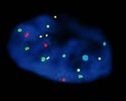
It is early morning, 6:30 a.m. I sit in a dark room, staticky music from WXRT playing on the beat up radio in the corner. The setting is ripe for meditating, but my meditation consists of peering through a large, clunky, microscope. I see glowing blobs of blue, shiny pairs of dots reds and yellows, greens and aquas. I make notes on a score sheet. I am without a rod and reel, there is no lake or river nearby, but I am on my morning FISHing trip.

I’ve talked about prostate cancer and looking at biopsies a few times here, but we actually process more specimens related to bladder cancer. Sometimes we get small biopsy samples, taken by the urologist during a cystoscopy procedure, but more often it’s the Yellow River that provides the material for us to analyze.

Most people don’t realize that urine contains a myriad of cells that drift off the inner lining of the bladder into your pee. Our job in the lab is to look at those cells and determine if any of them are suggestive of cancer in the bladder. How do we do it? We have a couple of different methods.

Our simplest test is called cytology. Our great lab team first centrifuges and then processes the urine specimen until a thin layer of cells covers a glass slide. Those cells are then stained with the Papanicolaou stain, and yes that is the same one used in a traditional Pap smear. Several pairs of eyes look at the stained cells, and any significantly abnormal ones are noted and reported. We do about 50 of those cases a day.

But my early mornings are spent in the dark room FISHing. FISH, which stands for Fluorescent In-Situ Hybridization, is a technique in which a glass slide covered with cells is stained with probes that mark specific chromosomes in each cell. Each probe is formulated to light up a different color when examined in the dark under ultraviolet light. Our special microscope is equipped with a UV light and four different wavelength filters. lab fishWe examine each cell, and by switching filters are able to count four different chromosomes. Two of a kind is normal, when a cell has an increased number of chromosomes, we start to worry.

Our system is automated. It is linked to a computer that can do the initial screening of the cells and then take digital photographs for the pathologists to examine. But I like to get into the FISH room in the morning, turn off the lights, and look at those cells myself.  Peacefully floating down the FISH river is a great way to start the day!

Type your email address in the box and click the “create subscription” button. My list is completely spam free, and you can opt out at any time.

 

No “Lovie” for Our Lab

illinoisWe’re Loyal to You Illinois…

…We’re “Orange and Blue” Illinois

The University of Illinois have some big news. Former Chicago Bears Head Coach Lovie Smith is the new Head Coach of the Fighting Illini football team. This really draws statewide attention to the school. What a tremendous boost for the team’s biggest challenge, recruitment.

And recruitment is tough. Now, I’m not talking about going to a high school students homes and convincing the child and his parents that he wants to battle on the offensive line in Champaign for the next four years. I’m talking about the battle we face in the lab every time we look for a technician or technologist with the special training and certification we require. There are a lot of laboratories in the Chicago area, and we all look to the same pool of potential employees. It is not a big pool, and there aren’t that many new people flowing through the spigot into that pool.

What makes a good place to work? Of course salary is important. Beyond that, one of our supervisors did some research and identified perks such as in house yoga, professional on-site chefs and variable work schedules. Regular demonstrations of appreciation also help. We haven’t tried the yoga or gourmet chefs here, though there are frequent celebratory lunches and birthday breakfasts. And we do try to offer a competitive salary, show respect to all of the staff, and allow some creativity within the bounds of all those detailed SOPs. But perhaps we need to do a better job with the pats on the back. And it is also important, though in a much longer term, to make sure there are always new students entering the lab pool, to help keep the spigot on the full-on position. We appreciate the schools and training programs that provide the needed education. And I speak at local career days, but not as often as I should.

What makes you want to stay with a company, or look for a new one? Will the place with the biggest budget always win out? How do we succeed in a field with a limited pool of qualified applicants? The challenge is always there for us. Maybe we DO need Lovie Smith to go knocking on some doors for us. And if that doesn’t work, does anyone know where I can find Mike Ditka?

On another note, I have an additional album title for our previous music trivia question.  “Stop Making Sense” is both a movie and a soundtrack album from David Byrne and the Talking Heads.  The phrase is from the song “Girlfriend is Better” that was performed live on the album.

To follow our blogs type your email address in the box and click the “create subscription” button. My list is completely spam free, and you can opt out at any time.


Don’t Believe the Hype! Diagnosing Cancer Not for the Birds.

dylanIt’s my work, he’d say…

…and I do it for pay

Bob Dylan

Hurricane, 1975

For most pathologists, it is more than a job, it is a calling. We are trusted to examine tissue from patients that we will most likely never seen or ever talk to, and provide a diagnosis that will often change their lives. The task requires a thorough knowledge of medicine, years of specialty training in pathology, and countless hours of microscope time learning the art, as well as the science, of our chosen profession. A well trained staff is also essential in making the correct evaluation, we are very much subject to “garbage in, garbage out.”

So most of us chuckled at bit at all the news reports last month about pigeons being trained to diagnose cancer. It seems that with a few pellets, any creature with good vision can be turned into a pathologist! Since progress on the new house is on short term (we hope) hiatus this weekend, I thought I would take the time to explain just how a pathologist actually renders a diagnosis, and how it gets back to the treating physician. There are many different types of labs, and the processes is different at each one, but I will focus on how we do things at my laboratory, an outpatient lab specializing in urology, and key in on prostate biopsies.

Our lab is part of the largest urology practice in the Chicago area.  About 60 urologists across the region identify men, who either because of physical examination findings or because of abnormal blood work, require prostate biopsies. I will spare you the messy details about how the biopsies are taken, but generally 12-16 areas in the prostate are sampled, with a needlelike core of tissue about a 20th of an inch thick and an inch and a half long taken from each site. Those samples are placed in jars filled with formalin, and yes, it smells just like what you remember from high school biology. The jars are carefully labelled and packaged for delivery, while patient information is  entered into the electronic health record we share with the urologist’s offices.

We use a courier service that specializes in handling medical specimens to bring the biopsies to the lab. That’s where our great processing team takes over. After verifying that all the information we have in the health record matches the specimen jars we have received, a description of the cores is dictated for our  report. They are then “cooked” in a microwave processor, embedded in paraffin wax (yes, the floors get slippery), and then cut into ultrathin sections which are placed on a labelled glass microscope slide and stained with colorful dyes. The sections of prostate turn blue and purple and pink.  Some of this work is automated, but much is done carefully, by hand, one slide at a time.

One of our four pathologists then looks at each slide under the microscope and formulates the diagnosis. How do we do it? We each  have an encyclopedic knowledge of what normal prostate looks like. We look for changes in the appearance of the stained tissues, subtle or obvious, that signal a change from normal to abnormal. We then mentally run through the myriad of possibilities that the abnormality could represent. Some of these are benign and of no significance, others indicate cancer, or a potential for future cancer. When we are uncertain, we can have additional slides made and use stains beyond the routine ones. Our diagnoses are then entered into the lab report. Before releasing the report, we have a final checkpoint. Each afternoon, our pathology group meets in my office. We gather around a video monitor connected to my microscope and review cases together. We do this for every case with a cancer diagnosis. Once we all agree, our completed report is signed and becomes part of the electronic health record, available to the urologist for action. In our lab, the whole process takes about 2 days from the time the urologist does the biopsy.

I love birds. Counting Crows, The Eagles and Flock of Seagulls are all music to my ears. But when it comes to making a diagnosis, leave the feathers behind. We may get paid in more than bird seed, but if you want the right answer, find a pathologist!

One last thing. An apology to all those who got tossed and turned by the broken links on our last post. If you missed it, you can find it here. This link will work. I promise!

As always, thanks for reading. Please comment, subscribe and share.

Type your email address in the box and click the “create subscription” button. My list is completely spam free, and you can opt out at any time.